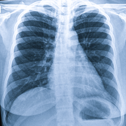
Canada: Radiologists should pay close attention to disease distribution and specific distinguishing imaging features when interpreting CT scans for fibrotic interstitial lung disease (ILD), a study published on March 17 in Radiology has revealed. The findings highlight how a more structured assessment of imaging characteristics may improve diagnostic accuracy in a group of conditions known for variable presentation and often poor prognosis. The study, led by Daniel-Costin Marinescu from the University of British Columbia, examined how individual CT features contribute to identifying radiologic patterns in fibrotic interstitial lung disease. Accurate classification of these patterns is critical, as it directly influences treatment strategies and clinical outcomes. The researchers emphasized that their findings could help clinicians, particularly non-experts, better understand the relative importance of specific imaging findings and support improvements in future diagnostic guidelines.Fibrotic ILD encompasses multiple subtypes, including conditions such as idiopathic pulmonary fibrosis and fibrotic hypersensitivity pneumonitis. Diagnosis largely depends on recognizing patterns on high-resolution CT scans, guided by criteria from the American Thoracic Society. However, these criteria have traditionally relied on expert consensus, and the contribution of individual imaging features to pattern recognition has not been clearly defined.To address this gap, the investigators conducted a secondary analysis of data from the Canadian Registry for Pulmonary Fibrosis (CARE-PF), including 1,498 patients evaluated between January 2021 and March 2022. Radiologic patterns were classified both according to established guidelines and the interpretations made by radiologists. The study applied statistical models, including multinomial analysis and receiver operating characteristic curves, to determine how well specific CT features differentiated between common ILD patterns such as usual interstitial pneumonia (UIP), fibrotic hypersensitivity pneumonitis (fHP), and nonspecific interstitial pneumonia (NSIP). The researchers reported the following findings:Certain CT imaging features were particularly useful in distinguishing between different ILD patterns.Increased honeycombing, along with reduced ground-glass opacity (GGO), was strongly associated with usual interstitial pneumonia (UIP).Areas of hypoattenuation were more indicative of fibrotic hypersensitivity pneumonitis (fHP).Nonspecific interstitial pneumonia (NSIP) patterns were characterized by higher levels of GGO and less honeycombing.Imaging features involving more than 10% of lung volume showed high specificity, often exceeding 90%.Discrepancies were observed between guideline-based classifications and radiologist interpretations.Mixed ground-glass patterns and central disease distribution led radiologists to diverge from guideline-defined UIP.Consolidation and peripheral or basal predominance contributed to disagreement in fHP classification.Honeycombing and reticulation influenced differences in NSIP categorization.Overall, the study found that radiologists tend to rely heavily on disease distribution and distinct imaging signs when identifying ILD patterns, with several features showing strong statistical associations with specific diagnoses. However, an accompanying editorial by experts from Johns Hopkins University noted that current fixed pattern classifications may not fully capture the complexity of ILD, as multiple patterns can coexist within the same patient.The authors concluded that a more nuanced, feature-based approach to CT interpretation could enhance diagnostic precision and better align imaging findings with clinical outcomes, paving the way for more personalized management of fibrotic ILD.Reference:Marinescu DC, Hague CJ, Muller NL, Murphy D, Churg A, Wright JL, Al-Arnawoot A, Bilawich AM, Bourgouin P, Cox G, Durand C, Elliot T, Ellis J, Fisher JH, Fladeland D, Grant-Orser A, Goobie GC, Guenther Z, Haider E, Hambly N, Huynh J, Johannson KA, Karjala G, Khalil N, Kolb M, Leipsic J, Lok S, MacIsaac S, McInnis M, Manganas H, Marcoux V, Mayo J, Morisset J, Scallan C, Sedlic T, Shapera S, Sun K, Tan V, Wong AW, Zheng B, Ryerson CJ. Association of CT Features with Radiologic Patterns in Interstitial Lung Disease: Multinomial Analysis in CARE-PF. Radiology. 2026 Mar;318(3):e251944. doi: 10.1148/radiol.251944. PMID: 41842670.
Original source: in