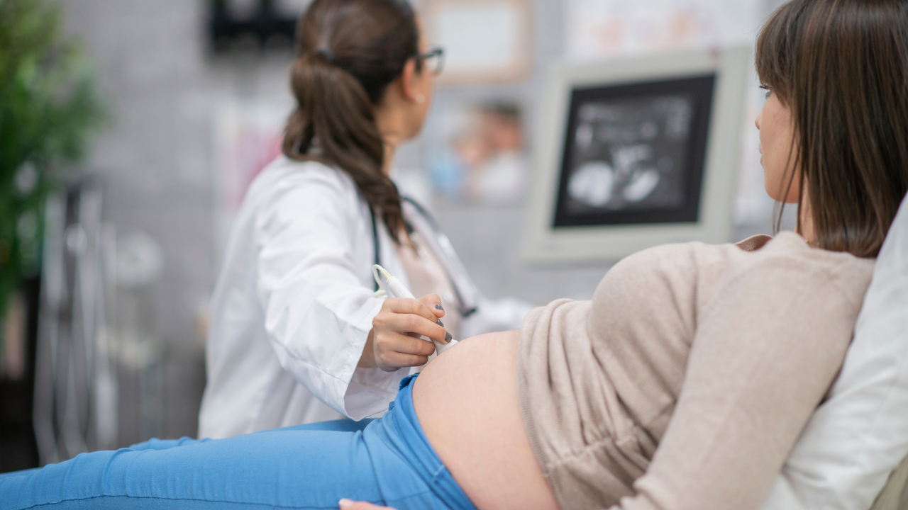
A team consisting of doctors successfully treated a rare and potentially life-threatening cervical ectopic pregnancy (implanted in the cervical canal instead of the uterus) in a 28-year-old woman experiencing her first pregnancy. Timely diagnosis, advanced interventional radiology, and a carefully planned multidisciplinary approach helped avert a major medical emergency. The patient recovered without complications, and her fertility was successfully preserved, allowing her to look forward to a healthy pregnancy and the possibility of becoming a mother in the future.Mrs Rashi Kumar (name changed), a resident of Mumbai, was thrilled when she discovered that she was expecting her first child. Like many first-time mothers, she eagerly began planning for the future and looked forward to the journey of pregnancy and motherhood. However, her joy was short-lived when a routine ultrasound at six weeks of pregnancy at a local sonography centre revealed an unexpected complication. The scan showed that the pregnancy had implanted in the cervical canal instead of the uterus, a rare and potentially life-threatening condition known as a cervical ectopic pregnancy. The diagnosis came as a complete shock to Rashi and her family, who had never heard of such a condition before. What had begun as a happy chapter in their lives suddenly turned into a medical emergency requiring immediate attention. Faced with uncertainty and fear, Rashi and her family sought specialised medical care at Wadia Hospitals.Dr Payal Lakhani, Additional Professor and unit head, MD, DNB (Obs and Gyn), Wadia Hospital, said, “The patient presented with a preliminary ultrasound report suggestive of a cervical ectopic pregnancy, a dangerous condition where the pregnancy implants within the cervix instead of the uterine cavity. Ectopic pregnancy is a pregnancy that is implanted at a site away from the uterus. These pregnancies do not grow into a normal pregnancy but cause the organ they are implanted into to rupture, leading to internal haemorrhage. It is a rare condition seen in approximately one in 10,000 to 15,000 pregnancies, increasing the risk of severe bleeding during evacuation. While the exact cause of cervical ectopic pregnancy is not known, factors such as previous uterine procedures, fertility treatments, or abnormalities in embryo implantation may contribute to its occurrence. After the patient was admitted to the Hospital, a detailed colour Doppler sonography was performed, which confirmed a very high blood supply to the pregnancy site. This increased the risk of severe, life-threatening bleeding if the pregnancy was removed without first reducing the blood flow. Serial Beta-hCG tests were done, which measure pregnancy hormone levels, and indicated that the ectopic pregnancy was continuing to grow. This was a cause for concern, as the risk of severe bleeding increases as the pregnancy progresses. We had to act quickly to prevent potentially serious complications and ensure the safest outcome for the patient, preserving her fertility. Therefore, an ultrasound-guided procedure was performed at Wadia Hospital on April 30, 2026, to safely manage the condition.”Dr Payal Lakhani further added, “We initially administered ultrasound-guided intrasac methotrexate therapy, which involves delivering medication directly into the pregnancy sac to stop its growth. This approach is often used to avoid surgery and preserve reproductive potential. Though there was fetal demise after the procedure, the blood supply to the cervix was very high. A decision was made to embolise the blood vessels supplying the pregnancy and then plan an evacuation procedure. An embolisation procedure followed by the evacuation of pregnancy was done on May 7th, 2026Dr Gireesh Warswdekar, Interventional Radiologist, Nowrosjee Wadia Maternity Hospital, said, “The biggest challenge in cervical ectopic pregnancy is the possibility of sudden and massive haemorrhage during treatment. Many such cases required emergency surgery and, in some instances, even resulted in the loss of the uterus. Advances in interventional radiology have improved the ability to manage these pregnancies safely. We performed an embolisation procedure, a minimally invasive technique in which tiny particles are introduced through a small catheter to block the blood vessels supplying the pregnancy. This reduced blood flow to the affected area and lowered the risk of severe bleeding. Once the blood supply was safely controlled, the pregnancy evacuation procedure was then carried out, where the pregnancy tissue was carefully removed from the cervical canal under medical guidance. The patient recovered smoothly and was discharged within 24 hours of the procedure without any complications on May 8th, 2026. This step-by-step approach allowed us to treat the condition safely and preserve the patient’s future fertility.”Expressing her gratitude, patient Mrs. Rashi Kumar (name changed) said, “Finding out that I was pregnant for the first time was one of the happiest moments of my life. I never imagined that the journey would take such an unexpected turn. When I was told that I had a rare condition that could lead to serious complications, I felt frightened and overwhelmed. Dr. Payal, Dr. Gireesh, and their team explained every step of the treatment, which gave me confidence during an incredibly difficult time. Although losing the pregnancy was heartbreaking, I am grateful that I still have the opportunity to become a mother in the future.”“Cases such as these underscore the importance of early diagnosis, multidisciplinary collaboration, and access to cutting-edge technology. We remain dedicated to providing the highest standards of care while continually advancing treatment options that improve outcomes and safeguard future reproductive health,” concluded Dr Minnie Bodhanwala, CEO of Bai Jerbai Wadia Hospital for Children.
Original source: in