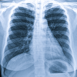
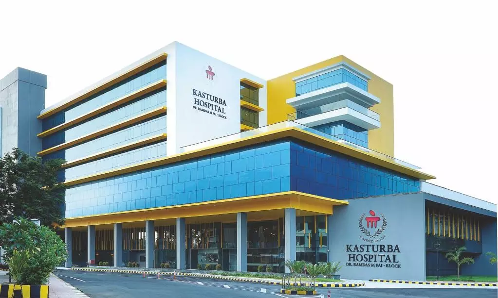
Manipal: A 60-year-old man who was admitted to Kasturba Hospital, Manipal, after vomiting blood was discharged within 48 hours after doctors controlled the life-threatening gastrointestinal bleeding using advanced endoscopic and endovascular approach, avoiding the need for surgery. According to the hospital, the patient was diagnosed with gastrointestinal bleeding caused by a fundal varix, a complication arising from chronic liver disease and portal hypertension. Doctors said bleeding due to liver disorders carries a high risk of mortality if not treated promptly. However, advances in endoscopic and interventional radiology techniques now enable such cases to be managed without surgery in many patients. An urgent upper gastrointestinal endoscopy performed by Dr Shiran Shetty, Gastroenterologist at Kasturba Hospital, revealed large fundal varices in the upper part of the stomach, one of which was actively bleeding. After the patient was resuscitated, the gastric varix was treated using EUS / endoscopy guided therapy to stop the ongoing bleed. Further endo-ultrasound, a newer technology, was used to detect the vein and shunt responsible for the bleeding. “Endoscopy today is not merely for making a diagnosis. The ultrasound at the tip of the scope let us pick out the exact vessel that had given way and treat it precisely, at a moment when the patient was critically unwell. Getting the bleeding under control quickly is what gives us the room to plan a lasting solution, instead of moving from one crisis to the next,” Dr Shetty said. Doctors found that a large abnormal channel, known as a gastrorenal shunt, had formed between the portal circulation and the left renal vein, continuously supplying blood to the gastric varices. They said sealing the bleeding vessel alone would not have been sufficient, as the bleeding was likely to recur if the shunt remained open. Following a joint discussion between the departments of Gastroenterology and Interventional Radiology, the patient underwent plug-assisted retrograde transvenous obliteration (PARTO), a minimally invasive procedure performed by the interventional radiology team led by Dr Harshith K. During the procedure, doctors accessed a vein through a single puncture and navigated a catheter to the abnormal shunt under X-ray guidance. A specially designed vascular plug was then placed at the opening of the shunt, blocking blood flow and preventing further filling of the gastric varices. A subsequent CT scan confirmed that the shunt had been completely sealed. The hospital said the patient experienced no further episodes of bleeding and was discharged within 48 hours of the procedure. A follow-up endoscopy by Dr Shiran Shetty confirmed that the varices had been successfully obliterated, with no evidence of active bleeding. The patient continues to do well during outpatient follow-up. MAHE Teaching Hospitals COO Dr Sudhakar Kantipudi said the case highlighted the benefits of multidisciplinary care.
Original source: in