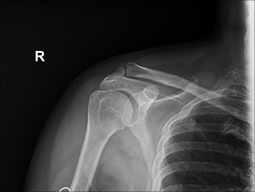
AP
shoulder X-ray to evaluate pain, injury, or discomfort in the shoulder joint. Common reasons include:
- Fractures: Identifying breaks in the humerus, scapula, or clavicle.
- Dislocations: Confirming if the humeral head is out of the glenoid cavity.
- Arthritis: Detecting degenerative changes in the joint.
- Bone abnormalities: Identifying bone spurs, infections, or tumors.
- Soft Tissue Calcification: Spotting calcific tendinitis


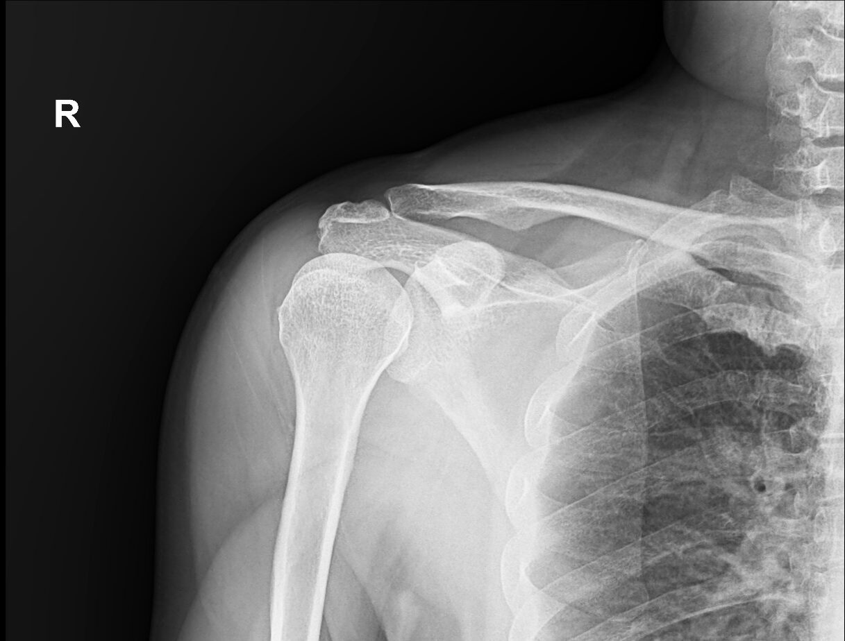
Internal rotation view
Radiographic projection used to visualize the shoulder joint, specifically highlighting the lesser tubercle of the humerus in profile medially
Transcapular view
also known as the Y view. assess suspected dislocations, scapula fractures, and degenerative changes.
- centering point: the level of the glenohumeral joint.
- exposure: 60-70 kVp, 10-20 mAs
- Distance: 100 cm
- grid: yes
Leave a Reply